Brain cells critical for mouse navigation found to be highly specialised
Researchers from Oxford University’s Department of Pharmacology have found that the ‘neural compass’ that enables mice to navigate is formed of specialised brain cells that respond in different ways to stimuli such as light and sound.
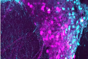
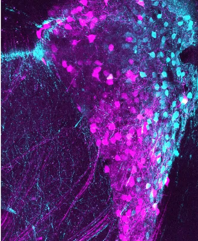
Calretinin-expressing cells in the mouse thalamus from the study (Image credit: Sara Hijazi).
The new study, published in Current Biology, reveals that these head direction (HD) cells are far more diverse than previously thought. Rather than simply encoding which direction an animal is facing, distinct groups of HD cells integrate information about light, sound, movement, and other sensory signals, helping the brain continuously update its internal sense of orientation. The findings provide new insight into how the brain combines information from the body and environment to support navigation and may ultimately help explain changes in spatial awareness seen in conditions such as Alzheimer's disease, autism, and schizophrenia.
To investigate how the brain's navigation system works, the researchers recorded the activity of individual HD cells in awake mice while monitoring the animals' orientation. They then labelled the same cells to examine their anatomy, connections, and molecular characteristics. This allowed the team to link each cell's activity with its physical structure and genetic identity, creating one of the most detailed maps yet of this key navigation circuit.
The researchers found that different HD cells responded in strikingly different ways to sensory stimuli. Some increased their activity when exposed to brief flashes of light, while others reduced their firing rates. Distinct subpopulations also responded differently to sounds and movement, suggesting that the brain's directional signals are shaped by information related to attention and arousal, as well as by orientation itself. Rather than acting as a single compass needle, the HD system appears to consist of multiple specialised channels that provide behaviourally relevant information about the surrounding environment.
Tim Viney, Associate Professor of Neuroscience and group leader in the Department of Pharmacology, University of Oxford, said: ‘Head direction cells are often described as the brain’s internal compass, but our findings show that they do much more than simply track which way an animal is facing. We found that these cells comprise distinct specialised subpopulations that combine directional information with signals about salient events in the environment, such as light, sound, and movement. This allows the brain’s navigation system to remain flexible and responsive to changing conditions. Understanding how these circuits work may also help us explain why disorders affecting this part of the brain can lead to altered attention, altered sensory processing, and spatial disorientation.’
The study also identified a previously unrecognised subpopulation of HD cells that express the protein calretinin. These cells had distinctive firing patterns and connectivity compared with other HD cells. The team further discovered an unusual calretinin-expressing cell type with twisted, corkscrew-like branches and a unique descending axon, which they named the "tortuosa" head direction cell after the corkscrew willow tree (Salix babylonica var. pekinensis 'Tortuosa').
The findings may have important implications beyond navigation. The brain region studied, the anterodorsal thalamic nucleus, is known to selectively express a number of genes associated with autism and schizophrenia, and previous research has linked dysfunction in this region to altered sensory processing and attention. In humans, the same region is particularly vulnerable to early neurodegeneration and is thought to contribute to the spatial disorientation often seen in the earliest stages of dementia. The researchers suggest that understanding how different HD cell types function could therefore provide new insights into both neurodevelopmental and neurodegenerative disorders.
The full paper, ‘Diversity and sensorimotor specialization of head direction cells in the mouse thalamus’, is published in Current Biology.


