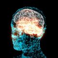
Blast trauma study suggests how to protect troops
An analysis of the brains of military personnel exposed to bomb blasts or concussive injuries, combined with experiments simulating the effect of blasts on the brain, suggests how soldiers could be better protected from improvised explosive devices (IEDs).
An international team led by Boston University researchers, including Professor Robin Cleveland of Oxford University, investigated the physical mechanisms that produce brain injury in soldiers subject to blast waves.
In their analysis the researchers found that brain tissue from military personnel who had been subjected to blast waves showed very similar damage to that observed in brain tissue from professional American football players. In American football players this damage has been associated with the progressive neuro-degenerative disease chronic traumatic encephalopathy (CTE), which is thought to induce depression, aggression, memory loss, and even suicide.
Laboratory experiments in mice demonstrated that exposure to a single blast equivalent to a typical IED does result in CTE and the long-term brain impairments that accompany the disease.
A report of the research is published in Science Translational Medicine.
Lee Goldstein of Boston University School of Medicine (BUSM) and Boston University College of Engineering, who co-led the study with Ann McKee of BUSM, said: ‘The neuropsychiatric symptoms of CTE that have previously been associated with athletes diagnosed with CTE could also be attributed to military personnel who were exposed to blast.’
Ann McKee of BUSM and the Neuropathology Service for VA New England Healthcare System said: ‘Our results showed that the neuropathology from blast exposure, concussive injury, or both were virtually indistinguishable from those with a history of repeat concussive injury.’ She added that the findings indicate that traumatic brain injury caused by different factors may trigger similar disease pathways in the brain.
In laboratory experiments in the US the researchers attempted to recreate the conditions at the heart of an IED explosion, where blast winds can reach a velocity of up to 330 miles per hour, to understand the physics involved and how brain injury might be prevented.
Using a mouse model in a shock tube, the dynamics of the animals’ head oscillations were tracked with a high-speed movie camera and very high accelerations were recorded. The brain tissue of the mice was examined and the morphology of the damage, and the damage to neurons associated with a specific protein that contributes to neuro-degenerative diseases, was the same as that observed in tissue from American football players and military personnel.
Professor Robin Cleveland of Oxford University’s Department of Engineering Science, a member of the team, said: ‘Animals subject to a single shock wave exhibited memory retention problems when given a task associated with finding their way through a maze. In contrast animals in which the head was immobilised did not exhibit tissue damage or memory problems.
‘The data suggests that it is the oscillation of the head that produces damage to tissue rather than the direct passage of the shock wave through the brain. The consequence is that to protect the head from injury the use of a helmet may not be the optimal strategy; rather a restraining device that reduces motion of the head and neck may be more effective.’
The research could also help to develop new ways of diagnosing blast-related brain trauma and better treatments and rehabilitation techniques for those who have suffered a blast or concussive injury.
A report of the research, entitled ‘Chronic Traumatic Encephalopathy in Blast-Exposed Military Veterans and a Blast Neurotrauma Mouse Model’, is published in Science Translational Medicine.
 Oxford scientists uncover how the brain resolves emotional ambiguity
 Expert Comment: In Claude We Trust? Evaluating the New Constitution
 Oxford tops QS World University Rankings in four subjects, named overall top for Humanities
 New study finds that stored sperm deteriorates across the animal kingdom
 Oldest genetic evidence for domestic dogs identified in Europe and Türkiye