Scientists 3D print human of the future
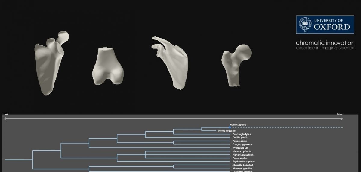
Interactive 3D models of human joints, showing how common medical complaints have arisen and how we are likely to evolve in the future, have been created at Oxford University.
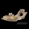
The researchers created 3D computer models of human joints by compiling 128 slice CT scans of bones from humans, early hominids, primates and dinosaurs. In all, they scanned 224 bone specimens.
By using 3D engineering and mathematical methods the group have produced 3D 'morphs' to plot changes in the shapes of species throughout the human lineage. This has provided new insights into the morphological trends associated with common orthopaedic complaints, such as anterior knee pain and shoulder pain.
Extrapolation of these trends has allowed 3D printing of possible future skeletal shapes as humans evolve.
Samples used in the study were from shoulders, hips and knees, and has enabled the researchers to make mathematical comparisons that could be used as planning tools for orthopaedic surgery. By comparing the modern and ancient samples, the team hopes to gain a better insight into the origins and solutions to common orthopaedic complaints.
Dr Paul Monk, who led the research at the Nuffield Department of Orthopaedics, Rheumatology and Musculoskeletal Sciences, said: 'Throughout our lineage we have been adapting the shape of our joints, which leads to a range of new challenges for orthopaedic surgeons. Recently there has been an increase in common problems such as anterior knee pain, and shoulder pain when reaching overhead, which led us to look at how joints originally came to look and function the way they do.
'These models may enable us to identify the root causes of many modern joint conditions, as well as enabling us to anticipate future problems that are likely to begin to appear based on lifestyle and genetic changes.
'Current trends reveal that the modern shapes of joint replacements won’t work in the future, meaning that we will need to re-think our approach for many common surgeries.'
The specimens scanned include amphibious reptiles (eg. Hellbender), dinosaurs, shrews, tupaiae, lemurs, primates, A. Afarensis (Lucy), H. Erectus (Turkana Boy) and H. Neaderthalis.
 Expert Comment: In Claude We Trust? Evaluating the New Constitution
 Oxford tops QS World University Rankings in four subjects, named overall top for Humanities
 New study finds that stored sperm deteriorates across the animal kingdom
 Oldest genetic evidence for domestic dogs identified in Europe and Türkiye
 Expert Comment: From frontier to feedback loop - Why space must become circular