
Image credit: Shutterstock
Diagnosing endometriosis without surgery
Tatjana Gibbons, a DPhil student at Oxford’s Nuffield Department of Women's & Reproductive Health, talks about research to develop a new 20-minute diagnostic test for endometriosis.
Around one in ten women will develop some form of endometriosis in their lives, a disease that affects more than 190 million women worldwide.
Yet despite its common occurrence, it typically takes eight years for a diagnosis – a figure that has shown no sign of improving in the past decade. The problem is that it typically involves several visits to the GP and hospital referrals, multiple scans and often surgery to diagnose.
Not only does this leave patients suffering for a significant period of time, but it can also require unpleasant and invasive procedures.
Aside from the pain, discomfort and worry about an ongoing undiagnosed condition, an important issue is its effect on fertility when left unchecked. A national questionnaire we conducted with over 1000 respondents with confirmed or suspected endometriosis found that 88% of respondents had experienced a delay in diagnosis, and 72% felt that an earlier diagnosis would have changed their life choices with regard to decisions such as fertility planning. With earlier diagnosis, it is possible to treat both the pain and discomfort as well as to begin to consider fertility options such as freezing eggs for the future.
The problem with diagnosing endometriosis is that small lesions in the pelvis are not detected on ultrasound and often need surgery to detect. This is why we are assessing alternative ways of looking inside the body without being invasive.
Our research group is conducting the DETECT (Detecting Endometriosis expressed inTEgrins using teChneTium-99m) imaging study, to investigate whether an experimental image marker (⁹⁹ᵐTc-maraciclatide) that binds to areas of inflammation can be used to visualise endometriosis using a 20-minute scan. In endometriosis, small parts of the lining of the uterus – the endometrium – are found in the fallopian tubes, ovaries or in the pelvis (for example, attached to bowel). The presence of the endometrium outside of the uterus leads to inflammation in the pelvis, which this test will aim to highlight.
If this is successful, it will give us a fast and effective way to identify any case of endometriosis, as well as assessing how serious the patient’s individual condition is and may even be able to distinguish between old and new disease.
Professor Christian Becker, who is Co-Director of the Endometriosis CaRe Centre in Oxford together with Professor Krina Zondervan, Head of Department at the Nuffield Department of Women’s and Reproductive Health, University of Oxford, will lead this initial study on women due to have planned surgery for suspected endometriosis.
Two to seven days before their operation, the participants will be invited for an imaging scan which will compare the suspected locations of disease detected on the scan and in surgery to confirm whether this imaging test matches what is found.
The beauty of this approach is that, if successful, it will provide a quick, painless and affordable option for tens of millions of women worldwide.
To read more about this research project and the partners involved, please visit https://www.wrh.ox.ac.uk/news/new-imaging-study-could-make-diagnosing-endometriosis-quicker-more-accurate-and-reduce-the-need-for-invasive-surgery
The study is being carried out in partnership with Serac Healthcare, a UK-based developer of breakthrough imaging technology.
 World Malaria Day 2024: an interview with Professor Philippe Guerin
 From health policies to clinical practice, research on mental and brain health influences many areas of public life
 From research to action: How the Young Lives project is helping to protect girls from child marriage
  Can we truly align AI with human values? - Q&A with Brian Christian
  Entering the quantum era
 Can AI be a force for inclusion?
 AI, automation in the home and its impact on women
 Inside an Oxford tutorial at the Museum of Natural History
 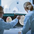 Oxford spinout Brainomix is revolutionising stroke care through AI
 Oxford’s first Astrophoria Foundation Year students share their experiences