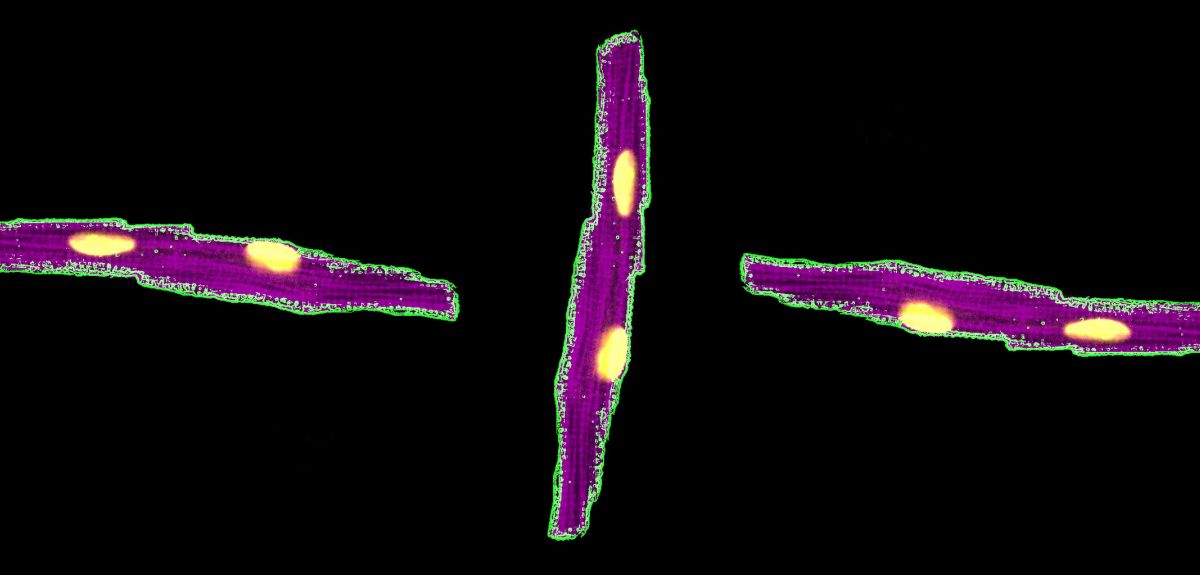
Image credit: Dr Matthew Daniels
Star Wars in a petri dish
A new method has been developed for observing the effects of drugs on heart cells under the microscope.
Researchers from Oxford University created the technique to test the effects of new or commonly used drugs on heart function, as well as exploring new ways to treat diabetes.
The new method involves flashing blue light onto the cell and watching how it responds by looking at it with a red light.
The technique involves inserting genes from blue-green algae and tropical coral into heart or pancreatic cells, which are created in the laboratory. The modified cells are introduced to a laser beam in a microscope, which the researchers named ‘the Death Star’, and the process is then filmed with a camera.
The laser beam turns the cells on and off, making them contract and relax to a fixed beat, which enables researchers to observe small changes that the drugs make in the way the cell responds.
For the past 60 years equivalent work has been conducted by capturing cells on a needle and then running an electric current through them.
Current experimental techniques are carried out on a cell by cell basis, which make them slow and inefficient, while the new method enables multiple cells to be used simultaneously. The new technique also enables cells to be controlled and observed remotely with light, rather than with direct contact.
Lead investigator, Dr Matthew Daniels from the Radcliffe Department of Medicine at Oxford, said: 'This research works using a simple microscope and a virus and means we no longer need to touch the cell or the dish. This allows hundreds of cells to be studied at the same time - so we have made the process a lot more efficient, and a lot easier to do.
'This will help us to identify drugs to treat diabetes and prevent sudden death, as we can detect helpful changes in pancreatic cells, and harmful changes in heart cells with this approach.
'It is a lot quicker and easier to do, meaning that work that usually takes three months and millions of cells can be conducted in three days on a handful of cells.'
The full paper, ‘Non-invasive phenotyping and drug testing in single cardiomyocytes or beta-cells by calcium imaging and optogenetics’, can be read in the journal Plos One.
 Researchers discover that ancient floods "rewrote" civilizations along the Yangtze River
 Statins do not cause the majority of side effects listed in package leaflets
 Are returning pumas putting Patagonian penguins at risk? New study reveals the likelihood
 Activism proves a stimulating topic at Sheldonian Series event
 New Oxford-led initiative launches to train future leaders in transformative technologies for pharmaceutical research